

Using Altair SimSolid for bone biomechanics – The University of Western Ontario
McFarlane Hand and Upper Limb Centre in London, Ontario is using Altair SimSolid to evaluate the biomechanics of bone stresses. Under the direction of Dr. Louis Ferreira, PhD, human bone specimens are CT scanned with a high-resolution scanner which preserves much of the internal trabecular bone's micro structure geometry. These models are imported into Altair SimSolid for simulated mechanical testing that matches the physical model. Altair SimSolid allows us to augment measurements from our experimental models that would otherwise be prohibitive or impossible to measure directly on the specimen.
Here are a few examples from our research.
[video width="2240" height="1440" mp4="https://www.simsolid.com/wp-content/uploads/2017/08/implant-2.mp4"][/video]
Here are a few examples from our research.
Shoulder joint implant
Patients with shoulder arthritis are often treated surgically by replacing the diseased joint with implants. This model shows the shoulder blade (scapula) with the shoulder socket replaced by a glenoid implant. Experimental loading conditions are simulated here through the arm implant components (not shown). This model allows us to simulate how different implant types influence bone stresses, which can influence longevity of the surgical procedure. Altair SimSolid allows us to test a high resolution bone model from a 60 micron resolution CT scan. Model created by Nikolas Knowles, MESc.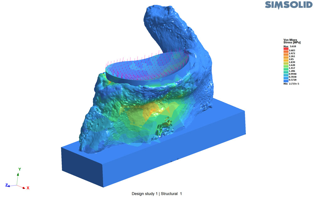
Image 1: Stresses on scapula bone with glenoid implant shown
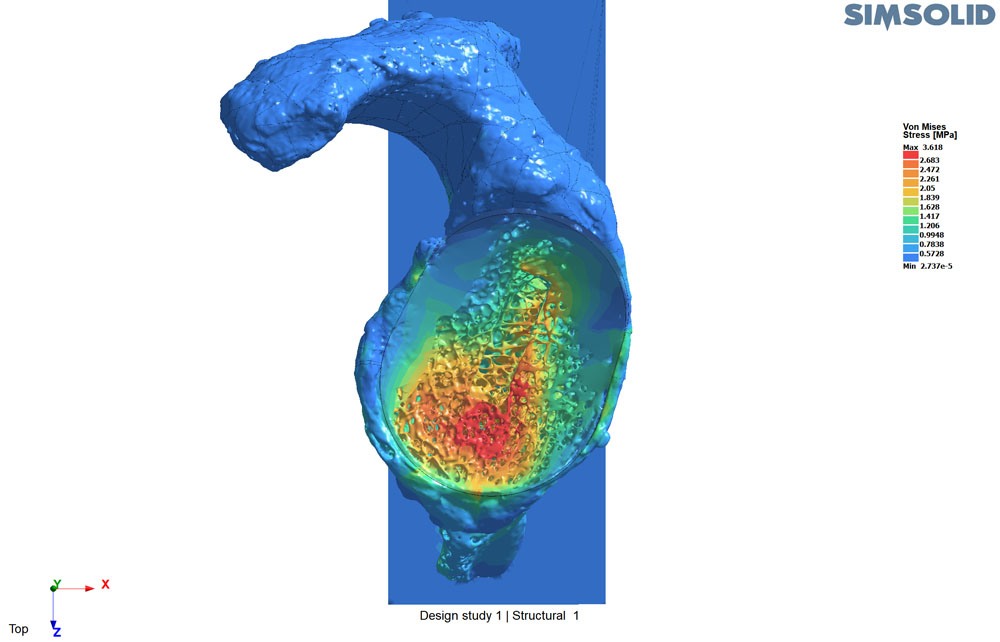
Image 2: Stresses on scapula bone with glenoid implant hidden
[video width="2240" height="1440" mp4="https://www.simsolid.com/wp-content/uploads/2017/08/implant-2.mp4"][/video]
Video 1: Stress animation on scapula bone with implant hidden
Human skull
An experimental model uses 3D printed piston mounts to recreate muscle lines of action on a cadaveric human head. Strain gauges on the bone surface measure bone strains generated by biting forces. This experiment is recreated in Altair SimSolid in order to measure bone strains in areas where strain gauges could not be placed on the specimen. The bone model was created from a CT scan. This model allows us to study the effects of injury and surgical implants like fracture repair plates and dental appliances. Model created by Kenneth Ip, BEsc and Nikolas Knowles, MESc.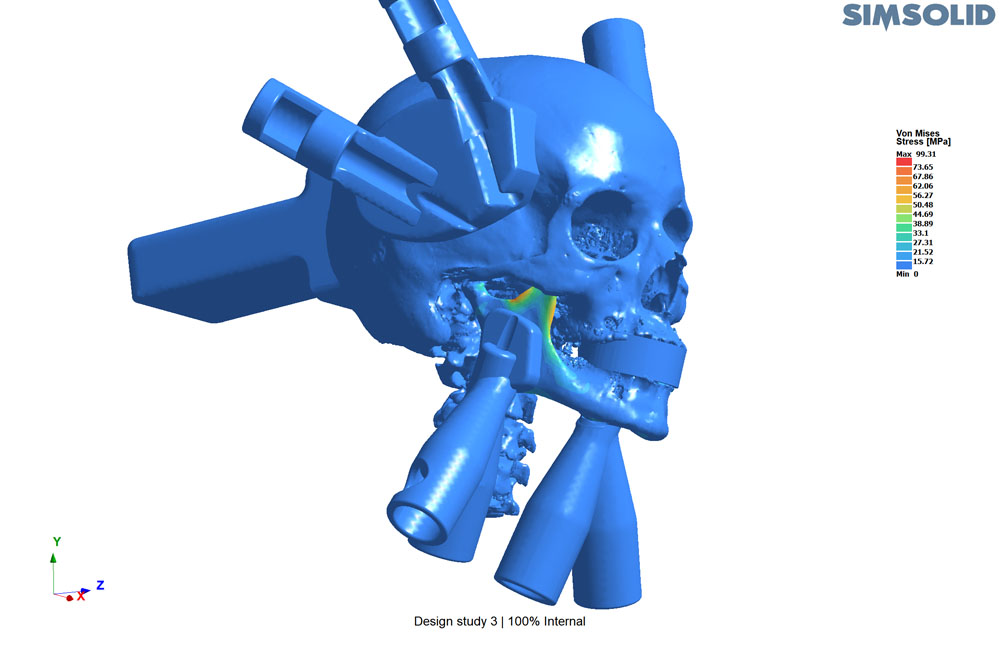
Image 3: Skull and jaw bone with 3D printed piston mounts
Concluding remarks
We have found Altair SimSolid to be an invaluable aid to our research work. It’s ability to analyze complex bone geometry is a capability that is not practical with other FEA methods. We look forward to using Altair SimSolid on more studies going forward.Louis M. Ferreira, PhD, Peng Associate Professor Department of Mechanical & Materials Engineering, Faculty of Engineering Department of Surgery, Schulich School of Medicine and Dentistry The University of Western Ontario